Shoulder Tendon And Ligament Anatomy / Anatomy Lab - Practical 1, Shoulder Ligament Model ... - Movement is therefore limited to flexion and extension.
byAdmin-
0
Shoulder Tendon And Ligament Anatomy / Anatomy Lab - Practical 1, Shoulder Ligament Model ... - Movement is therefore limited to flexion and extension.. The achilles tendon connects the heel to the calf muscle and is essential for running, jumping, and standing on the toes. Links the coracoid to the acromium and forms the. The distal joint between the tibia and fibula is an example of a. Shoulder muscles and shoulder tendons. Shoulder ligaments at louisiana state university.
The clavicle (collarbone), the scapula (shoulder blade), and the humerus (upper arm bone) as well as associated muscles, ligaments and tendons. Shoulder ligaments at louisiana state university. The anatomy of the provides the strength and functionality of the upper body. A joint capsule is a watertight sac that surrounds a joint. Bones in shoulder, ligaments of the shoulder joint, parts of the shoulder joint, shoulder anatomy, shoulder joints and muscles, shoulder structure anatomy, shoulder tendon anatomy, shoulder related posts of diagram of shoulder muscles and tendons.
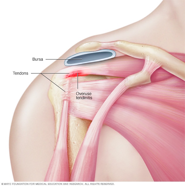
Shoulder joint - Mayo Clinic from www.mayoclinic.org The patellar tendon on the front of the knee is part of the quadriceps mechanism. It is a complex structure of bones, muscles and ligaments with the ability to lift weights and create enormous strength. Movement is therefore limited to flexion and extension. More about dental anatomy and periodontal ligaments you can find in the article about the anatomy of the teeth and this interesting video tutorial. The distal joint between the tibia and fibula is an example of a. Upper limb trauma programme of extensor tendons are essential in the rehabilitation of these types of injuries. Tendon and ligament injuries often go hand in hand with horses involved in vigorous athletic pursuits. This instability is countered by the strength of the rotator cuff muscles, tendons, ligaments, and the glenoid labrum.
The patellar tendon on the front of the knee is part of the quadriceps mechanism.
The anatomy of the provides the strength and functionality of the upper body. Anatomy of the shoulder these pictures of this page are about:shoulder tendons and ligaments anatomy. These ligaments are main source of stability for the shoulder. (3) a syndesmosis is a joint in which a ligament connects two bones, allowing for a little movement (amphiarthroses). Learn about their differences and the common injuries that affect them here. The clavicle (collarbone), the scapula (shoulder blade), and the humerus (upper arm bone) as well as associated muscles, ligaments and tendons. Muscles, tendons, and ligaments run along the surfaces of the feet, allowing the complex movements needed for motion and balance. Ligaments aid in joint stability during rest and movement and help prevent injury from hyperextension and hyperflexion (excessive movements). The shoulder can counteract an extreme impact but is also vulnerable to to a range of pathologies due to. Tendons and ligaments are bands of connective tissue that help stabilize the body and allow movement. Anatomy of the human body via wikimedia commons, public domain. The capsule, extensor tendon, and skin are very thin and lax dorsally, allowing for both phalanx bones to flex more. In the horse, lateral and medial movements of this joint are impossible due to the shape of the humeral head;
Ligaments and tendons are fibrous bands of connective tissue that attach to bone connecting two or more bones together and help stabilize joints. The capsule, extensor tendon, and skin are very thin and lax dorsally, allowing for both phalanx bones to flex more. Other smaller muscles and tendons surround the knee joint as well. The shoulder can counteract an extreme impact but is also vulnerable to to a range of pathologies due to. Once the ligaments, tendons, and muscles around the shoulder become loose or torn, dislocations can occur repeatedly.
Muscled Shoulder Joint Model - MedWest Medical Supplies from www.medwest.ca Start studying shoulder ligaments and tendons. Shoulder joint allows lifting, pushing and pulling by upper extremity. At the level of the pip joint, the. Anatomy of the shoulder these pictures of this page are about:shoulder tendons and ligaments anatomy. The human shoulder is made up of three bones: Tendons and ligaments commonly sustain injuries, which usually have similar symptoms and treatments. Tendons and ligaments are bands of connective tissue that help stabilize the body and allow movement. Mri may help your doctor identify injuries to the ligaments and tendons surrounding your shoulder joint.
At the level of the pip joint, the.
The patellar tendon on the front of the knee is part of the quadriceps mechanism. The shoulder joint is the articulation between the glenoid cavity of the scapula and the head of the humerus. Anatomy of the human body via wikimedia commons, public domain. Dr.g bhanu prakash animated medical videos. Learn about their differences and the common injuries that affect them here. Bones in shoulder, ligaments of the shoulder joint, parts of the shoulder joint, shoulder anatomy, shoulder joints and muscles, shoulder structure anatomy, shoulder tendon anatomy, shoulder related posts of diagram of shoulder muscles and tendons. Injury of tendons and ligaments remodel with scar formation with differences in themselves. Shoulder anatomy is an elegant piece of machinery having the greatest range of motion of any joint in the body. Roots, trunks, divisions, cords, branches, clinical anatomy. It is a complex structure of bones, muscles and ligaments with the ability to lift weights and create enormous strength. Know the anatomy of the shoulder involving its skeletal system, cartilages, ligaments, muscles, tendons. However, many tendon and ligament injuries can be avoided through proper conditioning and training regimens and by not pushing a horse beyond its limits in racing or other competitions. The shoulder joint (glenohumeral joint) is a ball and socket joint between the scapula and the humerus.
The shoulder can counteract an extreme impact but is also vulnerable to to a range of pathologies due to. Learn vocabulary, terms and more with flashcards, games and other study tools. (3) a syndesmosis is a joint in which a ligament connects two bones, allowing for a little movement (amphiarthroses). Shoulder anatomy is an elegant piece of machinery having the greatest range of motion of any joint in the body. These ligaments are main source of stability for the shoulder.
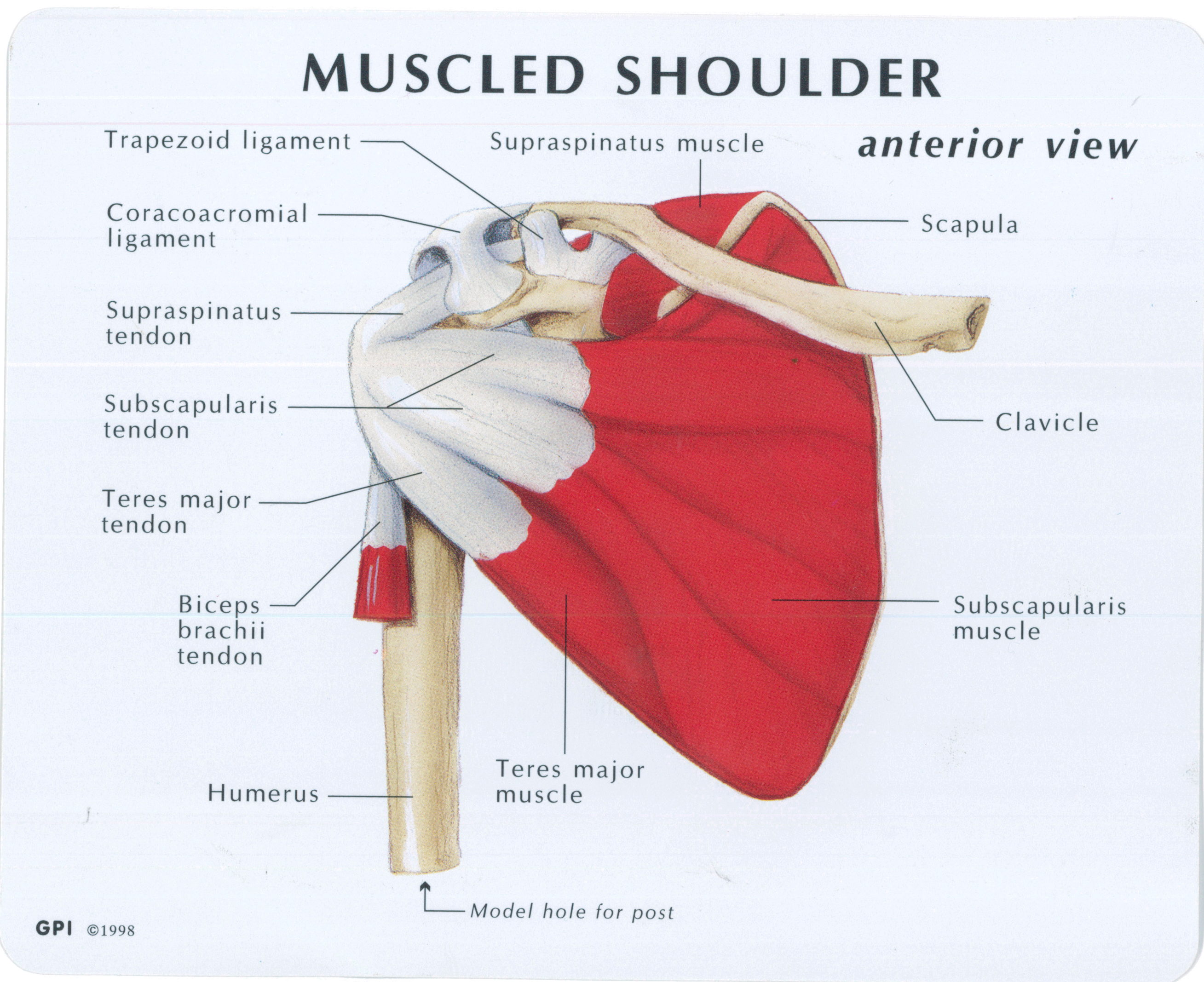
MRI Musculo-Skeletal Section: How to locate glenohumeral ... from 1.bp.blogspot.com The human shoulder is made up of three bones: Another condition that can affect ligaments is enthesitis, which is the inflammatory process within the entheses (the places where the tendons and ligaments. Scapholunate ligament and the lunotriquetral ligament are important intercarpal ligaments and disruption of either one can result in wrist instability. Transverse humeral ligament (thl) :holds the tendon of the long head of biceps brachii muscle in the groove between the greater and lesser tubercle on the humerus (intertubercular sulcus). Shoulder ligaments at louisiana state university. Roots, trunks, divisions, cords, branches, clinical anatomy. Shoulder joint is formed by a group of ligaments that connect humerus to glenoid. At the level of the pip joint, the.
Scapholunate ligament and the lunotriquetral ligament are important intercarpal ligaments and disruption of either one can result in wrist instability.
Tendon and ligament injuries often go hand in hand with horses involved in vigorous athletic pursuits. Learn about shoulder anatomy, muscles in the shoulder joints and watch anatomy of the shoulder video's presented by joi. Last update february 25, 2021. (3) a syndesmosis is a joint in which a ligament connects two bones, allowing for a little movement (amphiarthroses). Links the coracoid to the acromium and forms the. Anteriorly the subscapularis tendon is separated from the supraspinatus tendon by a gap, the rotator interval another important ligament, the coracoacromial ligament (cal). Ligaments aid in joint stability during rest and movement and help prevent injury from hyperextension and hyperflexion (excessive movements). Bones in shoulder, ligaments of the shoulder joint, parts of the shoulder joint, shoulder anatomy, shoulder joints and muscles, shoulder structure anatomy, shoulder tendon anatomy, shoulder related posts of diagram of shoulder muscles and tendons. Ligaments and tendons are fibrous bands of connective tissue that attach to bone connecting two or more bones together and help stabilize joints. Tendons and ligaments are complex structures and have different anatomical and dynamic properties. Muscles allow us to move by pulling on bones. The anatomy of the provides the strength and functionality of the upper body. Tendons and ligaments are bands of connective tissue that help stabilize the body and allow movement.
Although scarring depends on the quality and quantity of the injured tissues, it can be shoulder tendon anatomy. Related online courses on physioplus.